Regular screening mammograms hold a significant place in preventive care for women. By capturing detailed images of breast tissue with low-dose X-rays, these exams can reveal subtle changes that might not be noticed during a physical exam. They are utilized as a valuable resource for monitoring breast health over time.
What Is a Mammogram?
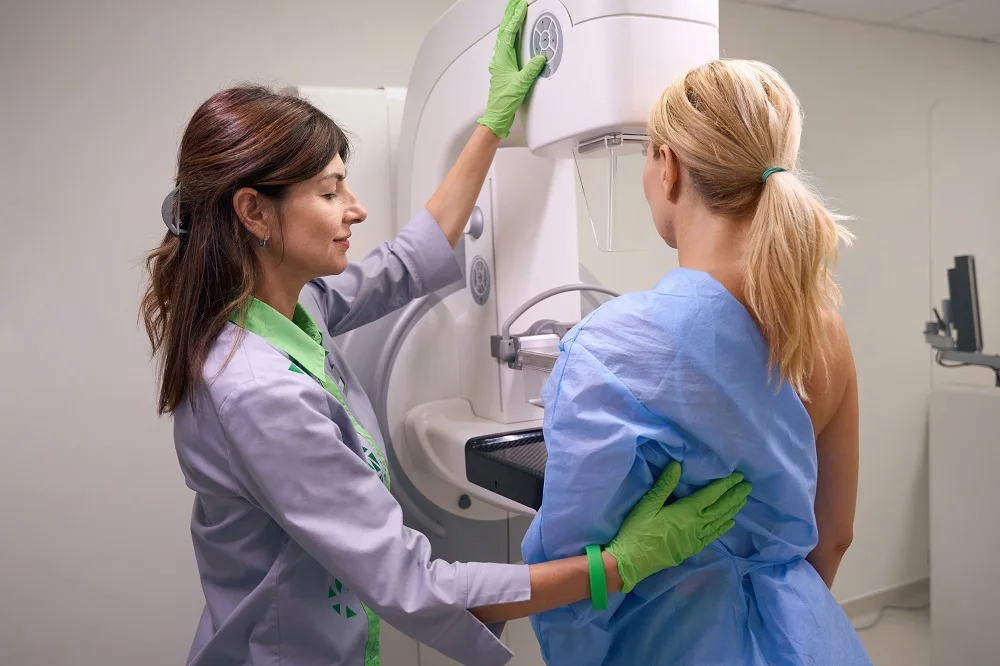
A mammogram is a low-dose X-ray of the breast that creates images of internal tissue. During the exam, a technologist positions the breast on a flat surface and applies brief compression to spread the tissue. This compression helps produce clear images and reduces the amount of radiation needed. The process typically involves several images from different angles and takes only a few minutes per breast. Screening mammograms are used when there are no breast symptoms to check for changes over time. Diagnostic mammograms are used when a specific area needs closer review, such as after a screening result or when symptoms are present.
Why Are They Necessary?
Breast tissue changes over time. Screening mammograms create a record that can be compared as time goes on. This comparison helps identify new findings or patterns that warrant a closer look. When changes are detected earlier, treatment options may be broader and outcomes may be more favorable. Mammograms can reveal small distortions that may not be felt during a clinical exam or self-exam. Detecting these changes at an earlier stage often allows for less intensive therapies. A smaller abnormality may be treated with more targeted approaches compared with changes found later.
When Should You Receive One?
Age, family history, genetic factors, prior breast biopsies, and breast density all influence timing and frequency. Some guidelines suggest starting routine screening in the early 40s, and intervals may range from annual to biennial. Your clinician can help tailor a plan that aligns with your values and risk.
If you have a strong family history of breast or ovarian cancer, or known genetic variants linked to higher risk, your timeline may differ. You may also discuss supplemental imaging, such as ultrasound or MRI, particularly if you have dense breast tissue. Dense tissue can make it harder to see certain changes on standard mammograms, so additional tools may be part of a personalized approach.
Practical steps can help you prepare, including:
- Gather Prior Imaging: Bringing previous mammograms or confirming they are available lets experts compare results.
- Share Your History: Inform your technologist and clinician about surgeries, hormone use, or symptoms like new lumps or nipple changes.
- Ask About Results: Clarify how and when you will receive findings, and whom to contact with questions.
If a screening result calls for extra views or an ultrasound, it does not mean there is a diagnosis. Most callbacks resolve with benign explanations. The key is to complete the recommended follow-up so you have clear answers.
Speak to Your Doctor
Regular screening mammograms are one tool within a broader breast health plan. The right schedule depends on your age, medical history, and personal preferences. A brief conversation with your clinician can help you select an approach that fits your needs and address questions about access.
- crypto30x com zeus Review: Is It the Best Platform for U.S. Crypto Traders in 2025?
- Super Scatter Juara100.org Medal: The Ultimate Guide to Winning Big in Online Gaming
- Ziuqyazhmizz: Ancient Slavic Practice Explained – Meaning, Benefits & Daily Life Guide
- Zaxtexporoz: A Simple Guide to Xcer Tools, Digital Trends, and Smart Solutions
- Casîo: A Symbol of Innovation, Durability, and Global Trust